MED-EL
Published Oct 31, 2013 | Last Update Jul 30, 2024
Pictures of the Ear: Deep in the Cochlea

You gave a great response to our 5 Amazing Facts About the Cochlea post, so we wanted to go a little further and show you some more real-life pictures of the ear and the parts responsible for hearing.
Let’s start with a picture of the whole inner ear, which is the part of the ear that contains the cochlea. You can see it in the picture at the top of this page. (That photo is courtesy of Helge Rask-Andersen, MD, Uppsala University Hospital, Uppsala, Sweden)
It’s is a plastic model of a real inner ear. The spiral-shaped structure on the left side is the cochlea, which contains the nerve structures that detect acoustic vibrations and turn them into the electrical information that the brain recognizes as sound.
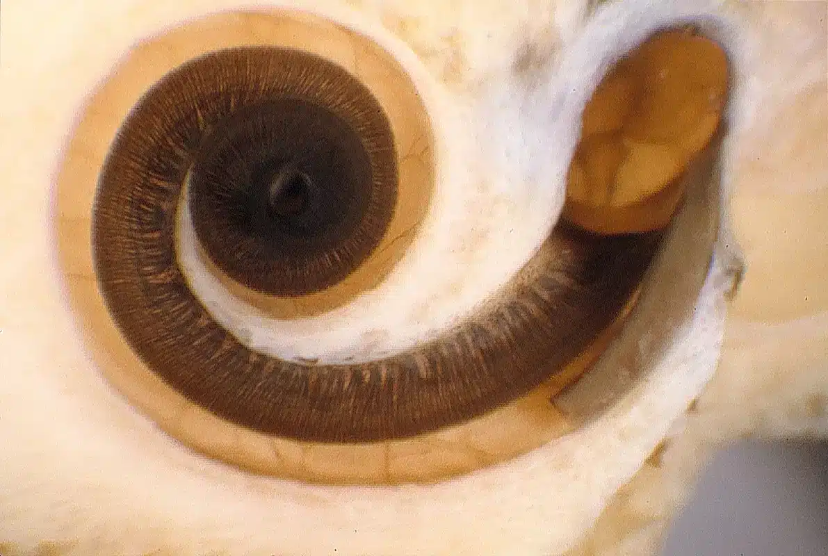
Now, let’s take a closer look inside the cochlea:
pictures of the ear deep into the cochlea1
Photo courtesy of C. G. Wright, Ph. D., UT Southwestern Medical Center, Dallas, USA.
This is a cochlea that has been opened so that you can see the nerve cells inside: each of the little lines that you see within the cochlea is a nerve cell. They are positioned all the way from the base (the top part in this image) to the apex (the innermost part of the spiral). Different parts of the cochlea respond to different frequencies of sound, from the very high frequencies (in the base) to the very low frequencies (in the apex).
But the cochlea isn’t a two-dimensional organ. As it turns it also curves up:
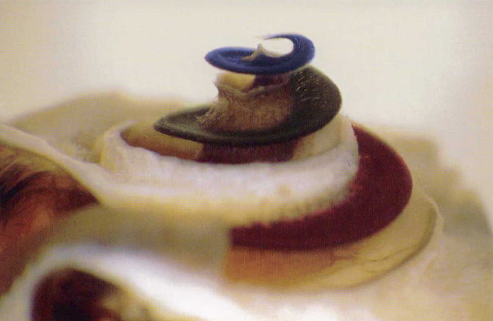
Photo courtesy of C. G. Wright, Ph. D., UT Southwestern Medical Center, Dallas, USA.
The innermost (and, in this picture, the uppermost) part of the spiral is the apex.
The cochlea is really small: from the very base to the apex, the average cochlea is only 31.5 mm long and only about 10 mm in diameter at its very widest point!
Here’s what it looks like when a cochlear implant electrode array is partially inserted:

pictures of the ear deep into the cochlea
Photo courtesy of Adrien Eshraghi MD, University of Miami Miller School of Medicine, Miami, USA.
It’s been cut in half so that you can see just how the electrode fits inside.
The electrode array consists of wave-shaped wires and metal contacts that are encased in silicon. The wires look silver because they’re made from of a mix of platinum and iridium, two metals that combine to create wires that are both strong and able to easily conduct electrical current.
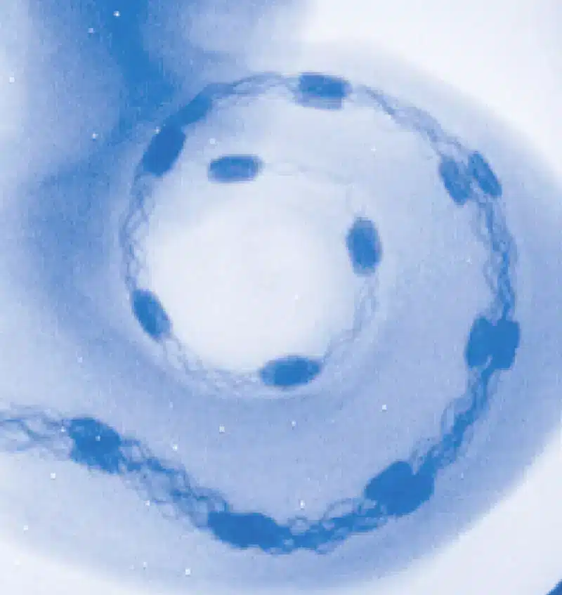
Photo courtesy of Univ.-Prof. Dr. med. Dr. h. c. Karl-Bernd Hüttenbrink, Uniklinik Köln, Cologne, Germany.
This is an x-ray of a cochlea with the electrode array inserted. You can see how the electrode array curves with the turns of the cochlea. Each of the dark spots in the electrode array is a metal contact, and it’s these contacts that transmit the electrical stimulation from the cochlear implant to the nerve cells of the cochlea.
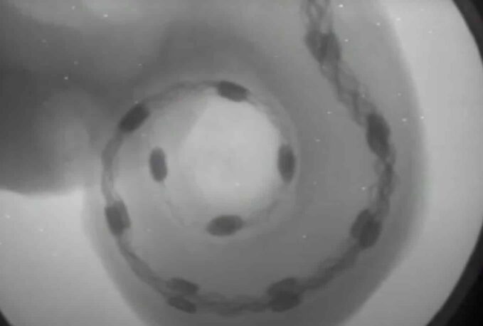
And if you’ve ever wondered what it looks like for a MED-EL electrode while it’s being inserted into a cochlea, check out the video below. The video is a fluoroscopy, which is a technique that uses X-rays to capture moving images in real time.
Find out more about cochlear implants and how they could help you hear.
Have you heard about SONNET 2? Discover how our latest cochlear implant audio processor is made for you.
Read more about how we design our cochlear implants to fit your individual ear.
References

MED-EL
Was this article helpful?
Thanks for your feedback.
Sign up for newsletter below for more.
Thanks for your feedback.
Please leave your message below.
Thanks for your message. We will reply as soon as possible.
Send us a message
Field is required
John Doe
Field is required
name@mail.com
Field is required
What do you think?
© MED-EL Medical Electronics. All rights reserved. The content on this website is for general informational purposes only and should not be taken as medical advice. Contact your doctor or hearing specialist to learn what type of hearing solution suits your specific needs. Not all products, features, or indications are approved in all countries.

MED-EL

MED-EL
